Gram Stain Lab Report
Aureus is a gram positive bacterium that when looked at under a microscope it appears to be a cluster of what. Antibiotic susceptibilities are only performed when appropriate CPT codes.
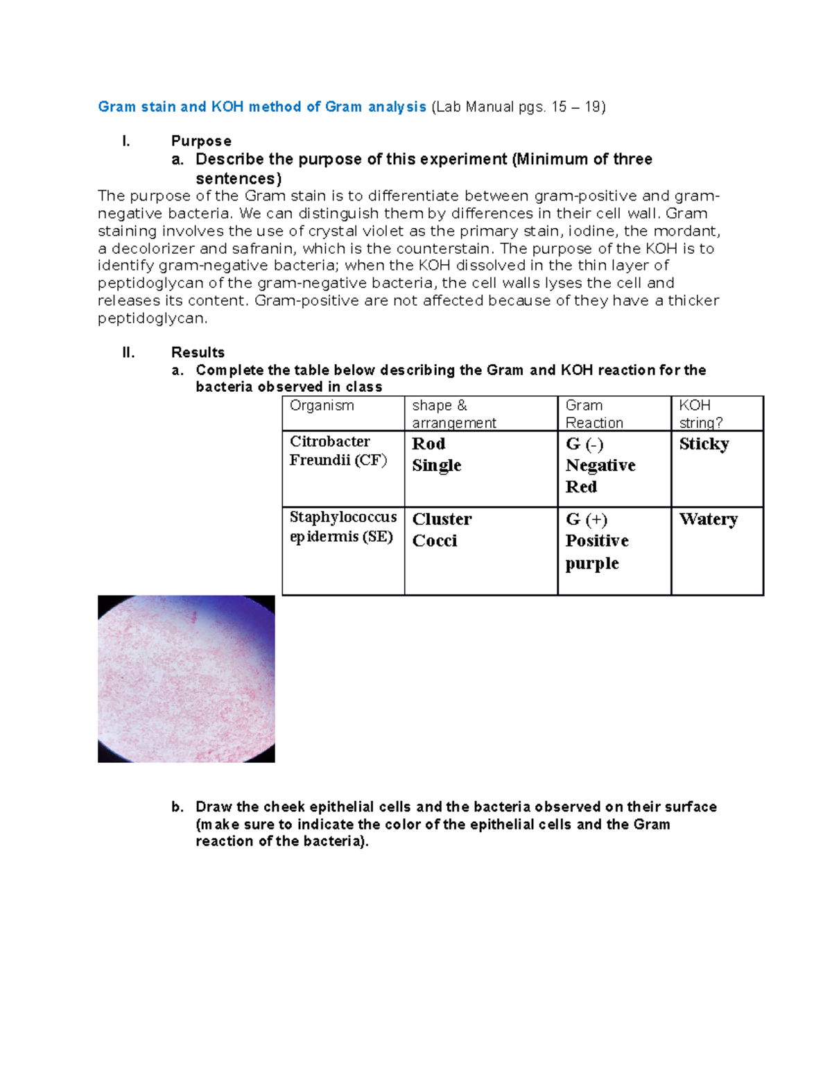
Gram Stain And Koh Method Of Gram Analysis Report Gram Stain And Koh Method Of Gram Analysis Lab Studocu
StaphsStreps Gram Enterics Gram- Motility.
. Example Lab Report of Staphylococcus aureus. Stab with a needle straight in and straight out of the center of the tube half. 87077 or 87140 or 87143 or 87147 or 87149.
Carbonic acid H 2 CO 3 CO 2 dissolved in blood and bicarbonate HCO 3- the predominant formBicarbonate is a negatively charged ion that is excreted and reabsorbed by your kidneys. Aerobic culture and Gram stain. Perform a Gram Stain to confirm culture purity from your subculture plate.
Using a sterile 5 mL pipette add 5mL of sterile saline to a sterile. Results obtained by culture without evaluation for contamination may be noncontributory or misleading. All sections including Intro Results Methods and Conclusion.
Dilutions in the range 10-1 110 to 10-8 range of dilutions can be restricted. Gram cfug of test material. A Gram stain is a way of detecting bacteria and is often performed on the same sample as a sputum culture test.
When doing a gram stain the results showed that the bacterium was Gram negative rods. Transport storage waste disposal Gram stain microscopy biochemistry measurements culture identification antimicrobial susceptibility testing typing methods serological or molecular. This lab should help give you the background information and techniques you will need to successfully perform general biochemical tests in order to help identify unknown.
The test report names the germ or germs that were found and the approximate amount of germs present. A gram stain was taken from a sample of the bacteria growing on the nutrient agar. The fungal spores shown are produced asexually The susceptibility of an organism to a set of antibiotics Resistant and more.
Our Gram ID cartridge technology is a rapid assay that measures differences in the cell walls of microbial isolates. GelRed 3X in water is ready-to-use for post-electrophoresis gel staining and is supplied in a 4L Cubitainer. For agarose gels we recommend using Original GelRed Nucleic Acid Gel Stain or GelGreen Nucleic Acid Gel Stain.
Gram-positive bacteria and gram-negative bacteriaThe name comes from the Danish bacteriologist Hans Christian Gram who developed the technique in 1884. Circular covering the whole dish irregular spreading out in a non-uniform pattern filamentous spreading out like roots towards the outer edge and rhizoid spreading out like branches with main segments. 87181 or 87184 or 87185 or 87186.
A bacterial wound culture is primarily used along with a Gram stain and other tests to help determine whether a wound is infected and to identify the bacteria causing the infection. If culture is positive identification will be performed at an additional charge CPT codes. Study with Quizlet and memorize flashcards containing terms like The pictured spores were most likely made by an Aspergillus mold.
Gram stain or Gram staining also called Grams method is a method of staining used to classify bacterial species into two large groups. The results showed the nutrient agar was only allowing one bacteria to grow. The gram stain turned out to be a positive rod gram stain.
If a culture reveals that a wound is infected susceptibility testing is done to determine which antibiotic will inhibit the growth of the bacteria causing the infection. Most commonly laboratories report a value for adjusted calcium also known as corrected calcium which is the measured calcium value adjusted for the albumin concentration. The information presented in this lab is from The Manual of Clinical Microbiology 8th Ed.
Common physical characteristics of bacteria colonies are listed and separated into 3 categories. In this section you should describe the results of both qualitative and quantitative methods of analysis. Choose the Right Stain for Your Application.
The bacterias form describes how they spread in a petri dish and can be. To confirm culture purity. Because we generally have no idea of how many bacteria are in a sample it is almost always necessary to prepare a dilution series to ensure that we obtain a dilution containing a reasonable number of bacteria to count.
All graphs and diagrams should have a number and title. SYTO 9 green fluorescent nucleic acid stain has been shown to stain live and dead Gram-positive and Gram-negative bacteria and it is a component of the LIVEDEAD BacLight Bacterial Viability Kits L-7007 L-7012 L-13152. Request forms report forms and other laboratory forms are all important components of the quality manual which documents the quality management system.
Conversely the the outer membrane of Gram negative bacteria is degraded and the thinner peptidoglycan layer of Gram negative cells is unable to retain the crystal violet-iodine complex and the color is lost. Gram staining differentiates bacteria by the chemical and physical properties of their. This is an image of conidia.
A Mannitol Salt Agar plate was obtained in order to test for mannitol fermentation. When you breathe you bring oxygen O 2 into your lungs and release carbon dioxide CO 2Carbon dioxide in your blood is present in three forms. The procedures are paraphrased from the National Committee for Clinical Laboratory Standards NCCLS 2000.
This complex is a larger molecule than the original crystal violet stain and iodine and is insoluble in water. As an automated stain-free assay the Gram ID cartridge eliminates technician variability that can occur in traditional Gram stain determination which involves multiple reagent steps and interpretation of results. This will be the bulk of your report.
Here is a good Example of an Unknown Lab Report in Microbiology. This is shown either numerically from 1 to 4 or with terms like rare few moderate or abundant. This is because abnormally high or low albumin concentrations can alter total calcium concentration independently of ionised active calcium concentration.
Designate a section of your report for your results. Make sure data is both graphed or diagrammed as well as described. 1 A Gram stain from a carefully collected specimen with neutrophils and lancet-shaped diplococci staining gram-positive which are intracellular or encapsulated can provide strong support to the clinical diagnosis of pneumococcal pneumonia.
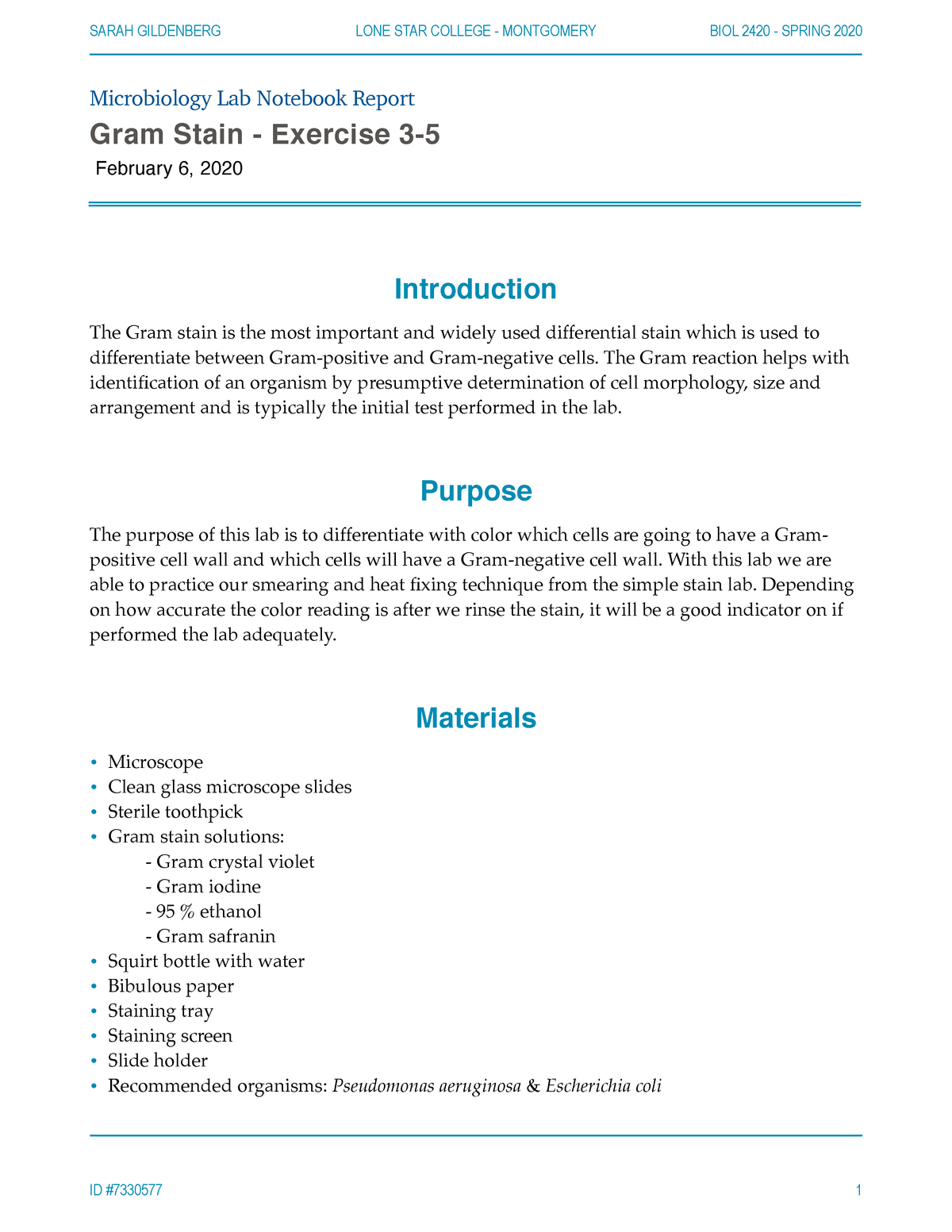
Complete Gram Stain Lab Microbiology Lab Notebook Report Gram Stain Exercise 3 February 6 2020 Studocu

Microbiology Gram Stain Lab Report Pdf Staining Histopathology

Pdf Mlt 415 Lab Report Gram Stain Techniques Muhamad Faizzudin Mohamad Zan Academia Edu
Gram Stain Lab Questions
No comments for "Gram Stain Lab Report"
Post a Comment